Ultrasound (US) and MR elastography (MRE) are both effective methods for assessing fibrosis in the liver. But which works better?

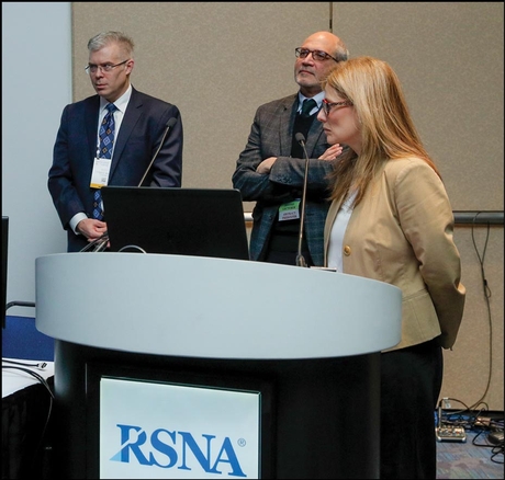
Left to right: Reeder, Sidhu, Kulik
It depends on the case, according to presenters at a Controversy Session Tuesday.
Fibrosis impairs liver function and is a risk factor for the development of cancer. It often arises from alcohol abuse, hepatitis C or nonalcoholic fatty liver disease. Left unchecked, fibrosis can advance to the much more serious condition of cirrhosis.
Elastography, an alternative to biopsy that noninvasively measures the stiffness of tissue in terms of units of pressure called kilopascals (kPa), has become a cornerstone of fibrosis assessment.
"Why are we doing elastography?" asked session co-presenter Paul S. Sidhu, MD, a professor of imaging sciences from King's College Hospital, London. "The answer is, we want to give an accurate assessment of liver diagnosis and what better way to do that than with numbers?"
Elastography can be performed with US or MRI. The US-based approach is more commonly used, having the advantage of wide availability and relatively low cost, Dr. Sidhu said.
Transient elastography, often referred to as FibroScan®, is perhaps the most well-established method. The US transducer transmits a low-amplitude signal to the liver, which induces an elastic shear wave that helps calculate stiffness. The stiffer the liver, the faster the shear wave propagates.
The transient approach is noninvasive, repeatable and samples a volume of liver 100 times greater than that of biopsy, Dr. Sidhu said. However, it is operator-dependent and its effectiveness is limited in obese patients and in the presence of ascites, or fluid accumulation.
ARFI is the Wave of the Future
Shear wave elastography and its close cousin, Acoustic Radiation Force Impulse (ARFI) imaging, represent the wave of the future, according to Dr. Sidhu.
"Shear wave is the easiest-to-perform scanning method as it's integrated into the ultrasound system," he said. "It's promising for detection of late stages of fibrosis and cirrhosis, but unproven in early fibrosis."
Research has shown that ARFI can tell with high confidence if a patient's liver is normal or cirrhotic. It is less effective at distinguishing the level of scarring, categorized on a scale from F1 for minimal to F4 for severe.
For that and other applications, MRE has significant value. The exam, performed by placing a passive transducer on the patient's right upper quadrant while they are in the scanner, offers a more advanced assessment of liver fibrosis, according to Scott B. Reeder, MD, PhD, from the University of Wisconsin School of Medicine and Public Health in Madison.
"The big difference with MR is you get more volumetric imaging of liver and a larger sampling," Dr. Reeder said. "In addition, MRI offers better wave penetration, technical feasibility and a higher technical success rate."
MRI can also identify other biomarkers of disease in the liver, something US is not equipped to do.
Dr. Reeder discussed several scenarios where MRE could play a role, including in obese patients, as a follow-up for patients with an abnormal US and in the screening of patients with higher pre-test probability of fibrosis. Treatment monitoring after a biopsy-proven diagnosis represents a particularly vital niche for MRE.
"Also, if you have a patient you're very worried about and there are contraindications to biopsy such as low platelet counts, then MRE would be a good option," he said.
From a hepatologist's perspective, elastography has been an enormously valuable tool for assessing fibrosis, according to Laura Kulik, MD, from the Feinberg School of Medicine at Northwestern Medicine in Chicago.
Dr. Kulik uses liver stiffness measurements in a variety of ways, such as in the diagnostic workup for cancer.
"We don't do as many biopsies as we did in the past," she said. "We do fibrosis measurements instead."

